




    Skip to main content
    Call Now: (770) 515-8770Schedule Today!
    Brain Conditions

    Epidural vs. Subdural Hematoma: A Complete Patient & Student Guide

    Dr. Marcus Stephens
    June 24, 2026
    8 min read

    Two Bleeds, Two Very Different Emergencies

    Both epidural hematomas (EDH) and subdural hematomas (SDH) are collections of blood inside the skull after a head injury — but they bleed from different vessels, behave differently on imaging, and demand very different surgical timelines.

    Understanding the difference matters because an epidural hematoma can kill within hours, while a subdural may evolve over days or weeks. This guide breaks down both in plain language for patients, families, and students.

    Quick Comparison

    | Feature | Epidural Hematoma (EDH) | Subdural Hematoma (SDH) | |---|---|---| | Bleeding source | Middle meningeal artery (arterial) | Bridging cortical veins (venous) | | Location | Between skull and dura | Between dura and arachnoid | | Onset speed | Minutes to hours | Hours to weeks (acute, subacute, chronic) | | Classic CT shape | Biconvex / lens-shaped | Crescent-shaped | | Crosses suture lines? | No (dura is firmly attached) | Yes | | Crosses midline? | No | Yes | | Typical patient | Young adult, temporal trauma | Elderly, anticoagulated, or alcohol use | | Classic history | "Lucid interval" then rapid decline | Gradual headache, confusion, weakness |

    The Anatomy Behind the Difference

    Epidural Hematoma — Arterial Bleed Above the Dura

    The middle meningeal artery runs in a groove along the inside of the temporal bone. A skull fracture across that groove — usually from a blunt blow to the side of the head — tears the artery. Because the bleeding is arterial and under high pressure, the dura is rapidly stripped from the skull, forming a biconvex (lens-shaped) mass on CT.

    The dura is firmly anchored at cranial suture lines, which is why an EDH does not cross sutures on imaging.

    Subdural Hematoma — Venous Bleed Below the Dura

    A subdural hematoma comes from tearing of bridging veins that travel from the cortex to the dural sinuses. These veins are thin-walled and low-pressure, so the bleed is slower and more diffuse. The blood spreads along the inner surface of the dura, producing the classic crescent shape that wraps the brain and freely crosses suture lines.

    In elderly patients, brain atrophy stretches these bridging veins, making them vulnerable to tearing from even minor head trauma — sometimes weeks before symptoms appear.

    Clinical Presentation

    Epidural Hematoma — The "Talk and Die" Pattern

    The textbook presentation is:

    1. Head trauma with brief loss of consciousness
    2. Lucid interval — the patient wakes up, talks, may even seem fine
    3. Rapid neurological deterioration: severe headache, vomiting, drowsiness, dilated pupil on the side of the bleed, weakness on the opposite side
    4. Coma and death within hours if untreated

    Not every EDH shows a lucid interval — but the hallmark is rapid arterial-pressure decline after a temporal-region head injury.

    Subdural Hematoma — Slower, Sneakier

    Acute SDH (within 72 hours) often presents like a severe head injury: depressed consciousness, focal weakness, pupillary changes.

    Chronic SDH (weeks after a minor fall or bump) is far more common in older adults and can mimic a stroke, dementia, or "just getting old":

    • Persistent or worsening headache
    • Confusion, memory changes, personality change
    • One-sided weakness or unsteady gait
    • Difficulty with speech

    Anticoagulants (warfarin, apixaban, rivaroxaban), antiplatelets (aspirin, clopidogrel), and alcohol use significantly raise the risk and severity.

    How Neurosurgeons Treat Each

    Epidural Hematoma — Emergency Craniotomy

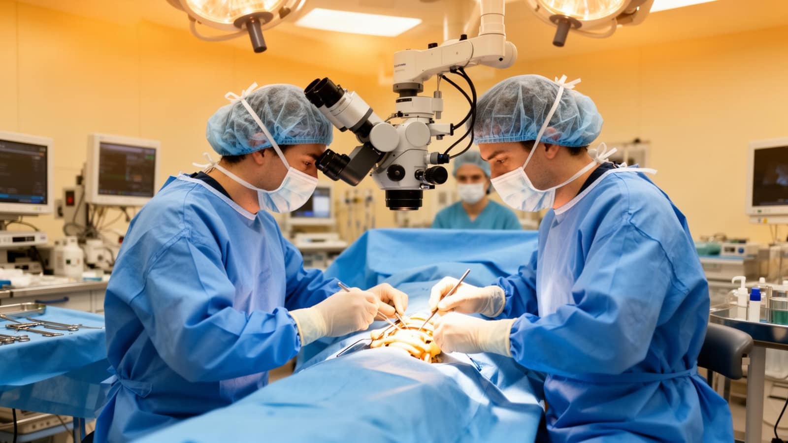
    A symptomatic EDH is a true neurosurgical emergency. Treatment is immediate craniotomy to:

    • Remove the blood clot
    • Find and stop the bleeding middle meningeal artery
    • Relieve pressure on the brain

    With prompt surgery, outcomes are often excellent because the underlying brain is frequently uninjured.

    Subdural Hematoma — Tailored to the Type

    • Acute SDH — usually craniotomy with clot evacuation; outcomes depend heavily on the underlying brain injury
    • Subacute / chronic SDH — often treated with burr-hole drainage, a smaller minimally invasive procedure where one or two small openings are made in the skull to wash out the liquefied clot
    • Small, asymptomatic SDH — may be observed with serial CT scans and reversal of any blood-thinning medications

    For a deeper look at the burr-hole approach, see our guide on subdural hematoma burr-hole surgery.

    When to Get Emergency Help

    Call 911 or go to the nearest emergency room after a head injury if you or a loved one develop:

    • Worsening headache that won't go away
    • Repeated vomiting
    • Drowsiness, confusion, or trouble waking up
    • Slurred speech, weakness, or numbness on one side
    • Unequal pupil size
    • A seizure
    • Any neurological change in someone on blood thinners after even a minor fall

    Long-Term Follow-Up at Nova Brain and Spine of Georgia

    Patients recovering from an EDH or SDH benefit from neurosurgical follow-up, repeat imaging, and a careful plan for any blood-thinning medications. At Brain and Spine Specialists of Georgia, our board-certified neurosurgeons — Dr. Jerry Walters II and Dr. Marcus Stephens — manage hematomas, traumatic brain injuries, and post-operative recovery across Hampton, Atlanta, and Middle Georgia.

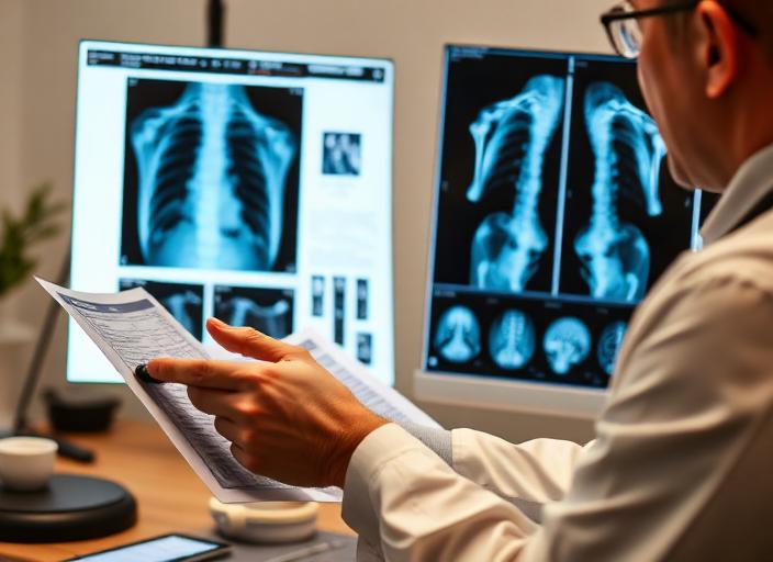
    If you have an MRI or CT showing a brain bleed, hematoma, or post-traumatic finding, we offer MRI reviews to help you understand your options.

    To schedule a neurosurgical consultation, call (770) 297-8133.

    Related Topics

    epidural hematoma
    subdural hematoma
    head injury
    traumatic brain injury
    neurosurgery

    Dr. Marcus Stephens

    Board-Certified Neurosurgeon, Skull Base & Cerebrovascular Specialist

    Board-certified neurosurgeon at Nova Brain and Spine of GA, providing expert care in Hampton, Georgia.

    Ready to Take the Next Step?

    Our board-certified neurosurgeons are here to help. Explore your options or schedule a consultation today.

    Related Articles

    Brain Conditions

    Brain Hematoma vs Brain Hemorrhage: Key Differences, Symptoms, and Treatment

    Hematoma and hemorrhage both involve bleeding in or around the brain, but they're not the same. Learn how neurosurgeons tell them apart and how each is treated.

    Dr. Marcus Stephens
    Jun 22, 2026
    7 min read
    Read full article
    Local Care

    Neurosurgeon in Hampton, GA — Herniated Disc Relief & Brain and Spine Care

    A neurosurgeon's guide for Hampton, Henry County residents: herniated disc relief, the conditions we treat most from your area, the drive from Hampton to our Hampton office, and how we coordinate with Piedmont Henry Hospital in Stockbridge.

    Dr. Jerry Walters II
    Jun 20, 2026
    7 min read
    Read full article
    Local Care

    Neurosurgeon in Lovejoy, GA — Sciatica & Lumbar Radiculopathy & Brain and Spine Care

    A neurosurgeon's guide for Lovejoy, Clayton County residents: sciatica relief, the conditions we treat most from your area, the drive from Lovejoy to our Hampton office, and how we coordinate with Southern Regional Medical Center in Riverdale.

    Dr. Marcus Stephens
    Jun 20, 2026
    7 min read
    Read full article