







Hematoma vs Hemorrhage: What's the Difference?
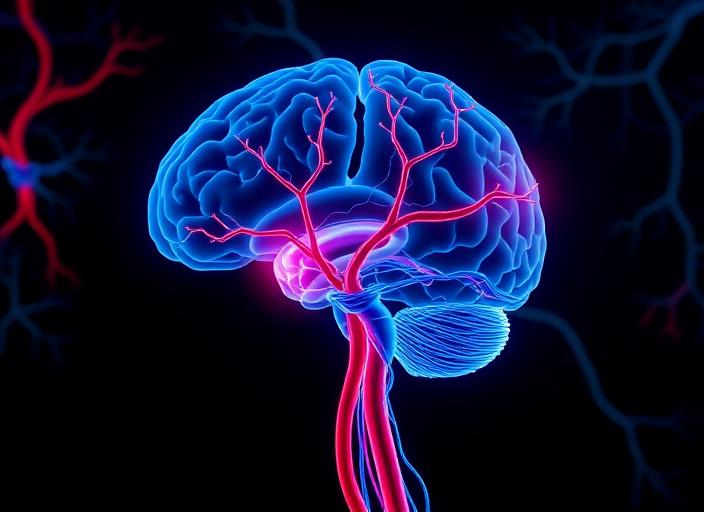
The terms brain hematoma and brain hemorrhage are often used interchangeably, but they describe different stages of the same underlying problem: bleeding in or around the brain.
- A hemorrhage is the active bleeding itself — blood escaping from a damaged vessel into brain tissue or the surrounding space.
- A hematoma is the collection of blood that forms once that bleeding pools and clots, often pressing on the brain.
Put simply: every hematoma starts as a hemorrhage, but not every hemorrhage becomes a large, organized hematoma.
Types of Brain Hemorrhage
Hemorrhages are classified by where the bleeding occurs:
- Intracerebral hemorrhage (ICH): bleeding inside the brain tissue, usually from chronic high blood pressure.
- Subarachnoid hemorrhage (SAH): bleeding into the space surrounding the brain, most often from a ruptured aneurysm.
- Intraventricular hemorrhage: bleeding into the fluid-filled ventricles of the brain.
Types of Brain Hematoma
Hematomas are classified by which layer of tissue the blood collects under:
- Epidural hematoma: between the skull and the dura — typically from a skull fracture tearing an artery. Symptoms can progress rapidly.
- Subdural hematoma: between the dura and the brain — often from torn bridging veins after a fall, especially in older adults or patients on blood thinners.
- Intraparenchymal hematoma: a clot that has formed within brain tissue after an intracerebral hemorrhage.
Common Symptoms
Both conditions can cause:
- Sudden, severe ("thunderclap") headache
- Nausea and vomiting
- Weakness or numbness on one side of the body
- Slurred speech or confusion
- Vision changes
- Loss of consciousness or seizures
Any of these symptoms after a head injury — or with no clear trigger — is a medical emergency. Call 911.
How Neurosurgeons Diagnose Them
Imaging is the key:
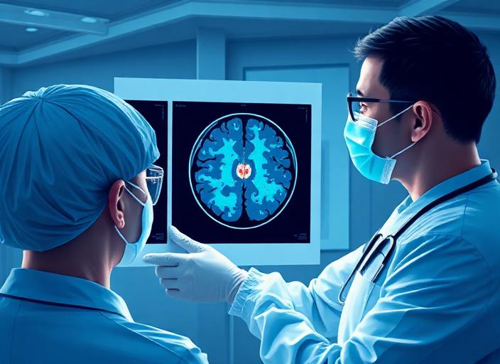
- Non-contrast CT scan — the first test in the ER; fresh blood shows up bright white.
- MRI — used to age the bleed and look at the surrounding brain.
- CT or catheter angiography — used when an aneurysm, AVM, or other vascular cause is suspected.
Treatment Differences
Hemorrhage management
- Aggressive blood pressure control
- Reversal of blood thinners when present
- Treating the underlying cause — for example, clipping or coiling a ruptured aneurysm in subarachnoid hemorrhage
Hematoma management
- Small, stable hematomas are often monitored with serial imaging in a neuro ICU.
- Large or expanding hematomas that compress the brain may need surgical evacuation — a craniotomy or burr-hole drainage depending on the type and location.
- Chronic subdural hematomas, common in older adults, frequently respond well to a small burr-hole procedure.
When to See a Neurosurgeon
You should be evaluated urgently if you have:
- A head injury followed by worsening headache, drowsiness, or vomiting
- Sudden neurological symptoms without a clear cause
- A known aneurysm, AVM, or prior brain bleed
- A new imaging finding of bleeding or a clot on the brain
At Nova Brain and Spine of Georgia, our board-certified neurosurgeons — including skull-base and cerebrovascular specialist Dr. Marcus Stephens — manage hemorrhages, hematomas, aneurysms, and traumatic brain injuries across Hampton, Atlanta, and Middle Georgia.
For an urgent neurosurgical evaluation, call (770) 297-8133.
Related Topics
Dr. Marcus Stephens
Board-Certified Neurosurgeon, Skull Base & Cerebrovascular Specialist
Board-certified neurosurgeon at Nova Brain and Spine of GA, providing expert care in Hampton, Georgia.
Ready to Take the Next Step?
Our board-certified neurosurgeons are here to help. Explore your options or schedule a consultation today.